
Abstract
Alopecia areata (AA) is a common autoimmune disease targeting the hair follicle and resulting in acute hair loss, partially or totally affecting the scalp and/or body. It usually affects young children and adults, causing severe psychological disorders and stress to the patients. There is still no definitive treatment for this disorder that can be persistent and recurrent. We present an interesting case of twins with genetic risk for autoimmune disorders who suffered from alopecia areata totalis. They had no significant response to conservative treatment with corticosteroids and were subsequently treated with platelet rich plasma (PRP) injections and showed noticeable and continuous hair growth simultaneously. We will discuss the potential of PRP as an alternative and trustworthy treatment for AA.
INTRODUCTION
AA is an autoimmune disease that targets the hair follicle, causing inflammation and, as a result, hair loss in several regions of the body. It is considered a genetic trait but may also be affected and/or triggered by environmental factors.1,2 PRP therapy is used to rejuvenate areas in need, as platelets have the ability to produce growth factors that locate and correct tissue damage. In the case of hair follicles, PRP activates follicular stem cells and improves the function of the hair follicle by prolonging the anagen phase. We previously reported a case of AA treated with PRP leading to very promising results and demonstrating a safe and effective alternative treatment in recalcitrant cases.6 Recently, we were presented with an even more intriguing case of AA with similar phenotype in dizygotic twins, who both also responded significantly to our PRP protocol.
Case Report
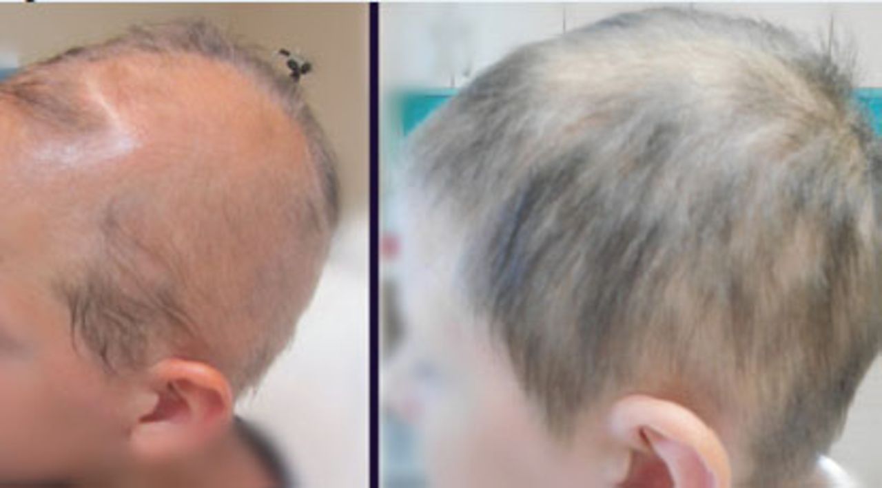
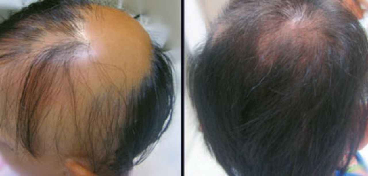
The 13-year-old male twins were referred to our clinic for hair loss that resulted in alopecia totalis. Four years ago, at the age of 9, one of the boys first presented with hair loss in his eyebrows and eyelashes. He remained stable for the next two years without any lesions on his scalp. According to his mother, he only used topical therapy based on corticosteroid lotions. Two years ago, the patient’s clinical appearance started progressing rapidly from the loss of round patches of hair at the back of his scalp to involve almost all scalp hair (Figure 1, left). He was then treated with oral prednisolone for 1.5 months and corticosteroid injections afterwards, resulting in only partial regrowth and poor duration. The other boy presented with clinical scalp involvement from the beginning, two years earlier than his brother (Figure 2, left). Their pattern of hair loss and progression of the disease appeared quite similarly. It is also of interest to note that although the twins’ medical histories revealed no comorbidities and no stress factors that could have contributed to the situation, their father reported suffering from vitiligo.


When the family consulted our clinic, they were introduced to our PRP protocol for AA, as the boys had already undergone all common treatment choices without noticing any significant and long-lasting improvement. The protocol involved 7 treatments of PRP with a one-month interval between treatments and the use of 5% minoxidil lotion once daily.
METHOD
The first step in PRP preparation is the collection of blood from the patient. We usually collected about 15-20ml from each of the twin brothers, depending on the session and the extent of the area to be treated. The vacutainers were then double centrifuged and, when ready, PRP was collected with a pipette from the tubes. After being activated with 0.05ml of 10% calcium chloride to each 1ml of PRP, 0.1ml/cm2 of the activated plasma was then injected into the patients’ scalp and/or eyebrows. To reinforce the results through trauma healing, at each treatment a Dermapen was also used for microneedling in some areas in combination with the injections.
RESULTS
The twins had a remarkable response to our treatment plan. Hair loss was diminished by the second session of PRP and hair regrowth started from the third session (Figure 1, right). Both patients presented with fully grown hair on the scalp and eyebrows one month after the last treatment, which is one of our quickest responses to PRP treatment for AA (Figure 2, right). Evaluation of the results was based on the patients’ photos and clinical presentation, as well as a trichoscopic examination before and after treatments. Follow-up also included a reexamination at 3 and 6 months post-treatment.
DISCUSSION
The occurrence of AA in members of the same family, especially twins, supports the theory that patients with AA are genetically predisposed to acquiring the disease sometime in their life.3 This assumption is supported by several twin studies, demonstrating the concordance rates in monozygotic as well as dizygotic twins.1,7 Rodriguez et al. studied the role of genes and the environment in the pathogenesis of AA, reporting concordance rates among 58 sets of twins.2 In the same study, AA was concordant in 42% of monozygotic and 10% of dizygotic twins, leading to the conclusion that the disease is not purely genetically determined because in such a case the rate in identical twins would be up to 100%.
Another interesting observation in our case is that the twins’ father had a personal history of vitiligo, another skin disorder of autoimmune etiology. There have been a few reports in literature suggesting that AA and vitiligo may share a similar pathogenesis and not be that different after all. The histopathological profile of both diseases consists of infiltrates of CD8+ T-cells in the epidermis and CD4+ T-cells in the dermis, highlighting the fact that compared to other autoimmune diseases (infiltration of T-cells, neutrophils, dendritic cells, and others), AA and vitiligo are less inflammatory.4 In addition, there have been reports of appearance of the two in family members due to shared genetic risk factors—direct overlap of associated genes and common gene categories (adaptive and innate immunity genes).5,8 Supporting the above, there have also been cases of patients with existence of AA and vitiligo at the same time or even at the same anatomical site in the skin, presenting with lesional overlap.4 Moreover, it is documented in other studies that patients with alopecia may present a higher risk for developing vitiligo compared to the general population.9,10
In our case, genetic risk certainly played a role in the appearance of AA in the twin brothers, and this hypothesis is further upheld by the fact that there was also another member in the family suffering from a similar autoimmune disease. Interestingly, our treatment plan proved remarkably successful given the background of the condition, enhancing the idea of PRP being one of the most promising therapies to date for AA. Although there have been many studies supporting the role of PRP in wound healing and teeth osteosynthesis as well as bone grafts, it is curious that published evidence about its efficacy in hair loss and certain types of alopecia appears quite poor.11 Our study resulted in the twins experiencing significant regrowth of scalp hair that started in 3 months and resulted in a major increase in their hair thickness and density.
CONCLUSION
This case reflects the successful results of the use of PRP in alopecia areata, even in cases with genetic predisposition. It may not yet be settled as an official treatment option for autoimmune hair disorders, but it consists a very promising as well as safe alternative choice for recalcitrant cases that have not previously responded to common treatments.
- Copyright © 2018 by The International Society of Hair Restoration Surgery
References
- 1.
- 2.
- 3.
- 4.
- 5.
- 6.
- 7.
- 8.
- 9.
- 10.
- 11.