


At the very first ISHRS meeting in Dallas, when I was just an infant in the great big world of hair transplantation absorbing as much knowledge as possible, Dr. Jung Chul Kim presented his fascinating data regarding follicular regeneration. Surprisingly, he demonstrated that hairs could grow without the papilla! When transected mid shaft horizontally, the upper half produced 40%, and the lower half 20%. This demonstrated that regenerative cells were not exclusively located in the papilla, and it was not necessary for reproduction of the follicle.1
Dr. Bobby Limmer in 1994 published his data indicating that the upper half produced new follicles 7.1% of the time, compared to 21.9% production with the lower half of the follicle. He observed that the caliber of most of these new follicles were of smaller.2
Two ideas were planted in my head from these two studies:
If the follicle could always be transected at the “perfect” level so that regenerative cells could always be present in the upper and the lower segment, theoretically one could double hair production.
Because the resultant hair produced from a transected follicle is smaller caliber, one could possibly find a use to further soften the frontal hairline for men and particularly women, also eyebrows and even eyelashes.
The first question was partially answered with 133% production from transected follicles, which I presented at the ISHRS meeting in September of 19983 and the second in a study performed at the Live Workshop in Orlando in the Spring of 1999, placing bisected follicles in the frontal hairline.4,5
With the above abbreviated history in mind and assistance of a grant from the ISHRS and the help of Drs. Jim Arnold, Marco Barusco, Michael Beehner, Jung Chul Kim, Jennifer Martinick, David Perez-Meza, and Craig Ziering, we set out to evaluate the following characteristics of single-hair follicular units bisected in the region of the bulge, and follicles transected through the papilla compared to controls:
Rate of growth
Pigmentation of resultant hair
Curl
Maintenance of implantation angle
Percent production
Time in tellogen effluvium
Extend the study for years to evaluate the above qualities in subsequent hair cycles
Study Design
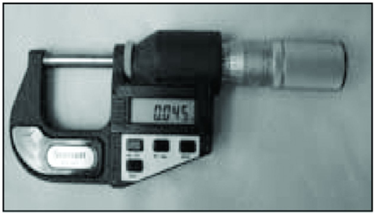
This study was initiated at the Orlando Live Surgery Workshop on March 2, 2000. Donor hair was removed from Dr. Mayer’s occipital area by Dr. Ziering. Subsequent microscopic dissection was performed with the 10× Meiji scope. Only those naturally occurring as “single” follicular units were selected. The hairs were 1–2cm long for accurate diameter measurements. These grafts were organized in three rows of 20 on chilled saline telfa pads by Dr. Martinick, and each was identified by row number and position in the row. She then measured the diameter of each hair with the Electronic Digital Starrett Micrometer (Figure 1).
Following the diameter measurement, Dr. Kim, with the 10× Meiji dissecting microscope, bisected 20 single follicular units at the level of the insertion of the arrector pili muscle in the area of the bulge. Dr. Kim also transected 20 single follicular units midway through the papilla returning them to their proper row and graft number.
Next, Dr. Beehner tattooed three vertical rows of 20 gray dots in each row on Mayer’s right upper anterior thigh. Row I was 20 control, intact single follicular units; Row II, the bisected follicles (upper segment medial to the dot, lower segment lateral to the dot); and Row III consisted of placing the 20 upper segments medial to the dots and the lower half of the papilla placed lateral to the dot.
A device created by Dr. Arnold to maintain and 18 gauge needle at 45 degrees downward was used to prepare all receptacle sites.


Periodically the following measurements were obtained:
Growth or no growth
Length in mm
Diameter in microns
Color
Curl characteristics
Skin evaluation for epidermal cysts, persistent erythema, etc.
Angle of exit at the dermal cutaneous junction
Results
Production
Row I Control: 12 of 20 = 60%
Row II Upper Segment: 10 of 20 = 50%
Row II Lower Segment: 6 of 20 = 30% Row III Upper Segment: 7 of 20 = 35%
Row III Lower Segment of Papilla: 0 of 20 = 0%
Growth Rate
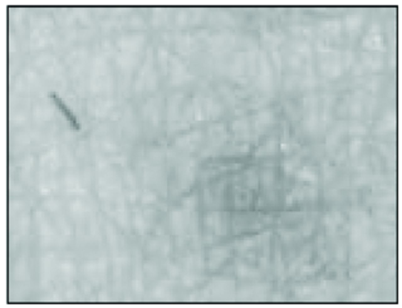
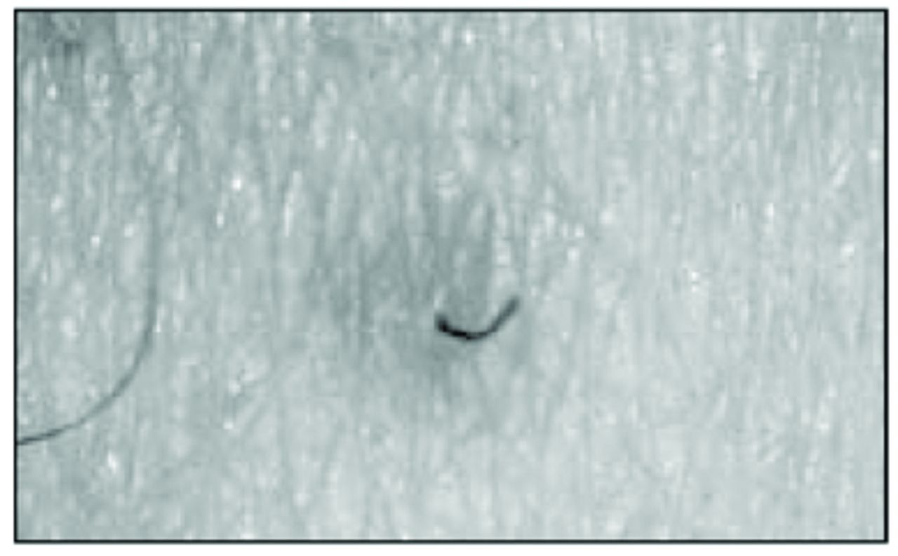
These measurements are markedly limited because of mechanical trauma to the hair follicle in the area of the upper thigh. Despite the use of multiple protective bandages and silk undergarments, most hairs were adversely affected by mechanical trauma (Figure 2).
Row I Control: Only hairs #6, 14, and 17appeared not to be traumatized. Hair #6 grew 53mm, #14 grew 35mm, and #17 grew 51mm. 53 + 35 + 51 = 46.3mm. Over 23 months = 46.3/23 = 2.02mm/month growth rate.
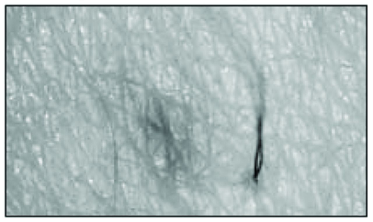
Row II Upper Segment: All were affected by trauma (Figure 2).
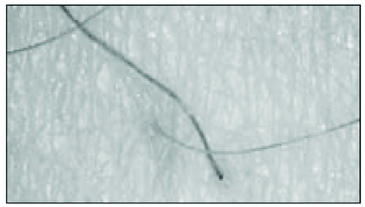
Row II Lower Segment: Only seemed to be unaffected by trauma (Figure 3). Hair #12 = 32mm; 32mm/23 months = 1.4mm/month
Example of two hairs from one follicle (Figure 9).
Row III: All less than 1mm due to trauma.
Diameter
Row I Control: Only 3 of the 12 hairs that grew were not broken off at the dermal cutaneous junction. Hair #6 = 46um, #14 = 64um, #17 = 57um.
Diameter difference pre- and posttransplant:
Hair #6: 57 − 46um = 11um
Hair #14: 64 − 64um = 0um
Hair #17: 60 − 57um = 3um
Average decrease in diameter 23 months transplant: 11 + 0 + 3/3 = 4.7um
Row II Upper Segment: Because all hairs appeared traumatized and had lengths less than 1cm, the Starrett Micrometer could not measure these. However, they appeared to be in the 25−35um diameter range.
Row II Lower Segment: Only one of the six hairs survived the mechanical trauma to grow to a measurable length.
Hair #12: 38um
Diameter difference pre- and posttransplant: 61um − 38um = 23um
Row III: Of the 35% production, none escaped the trauma to be sufficiently measured by the micrometer.
Color
Row I: Little or no change from the medium brown color.
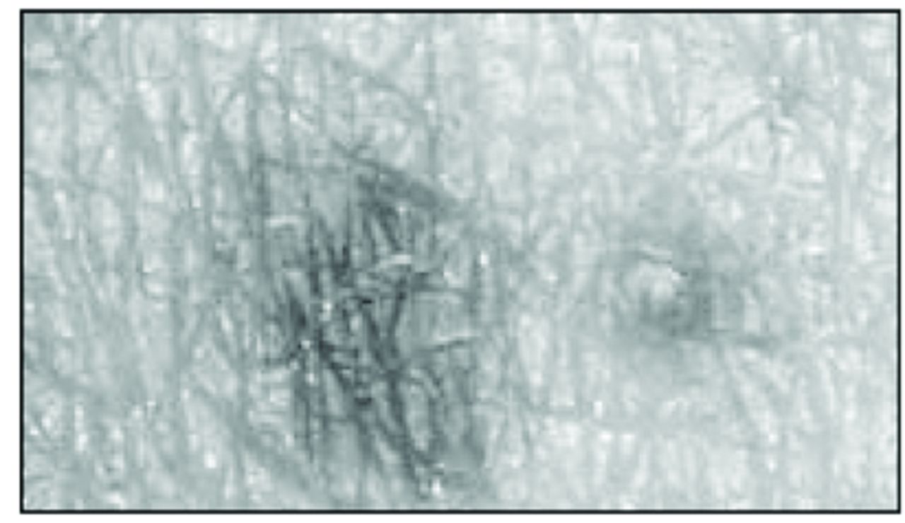
Row II and III: Most appeared lighter brown (Figure 4).
Curl Characteristics
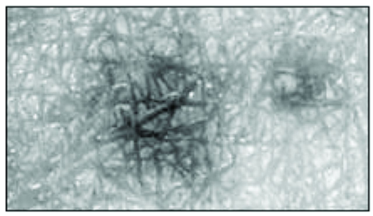
Row I Control: Increase in curl (Figure 5, hair #6 & #14)
Row II: Upper Segment: Length insufficient to determine Row II: Lower Segment: Hair #12 marked curl (Figure 6)
Row III: Length insufficient to determine
Skin Characteristics
Row I Control: Little or no perifollicular reaction
Row II: Upper Segment: Increased perifollicular inflammation
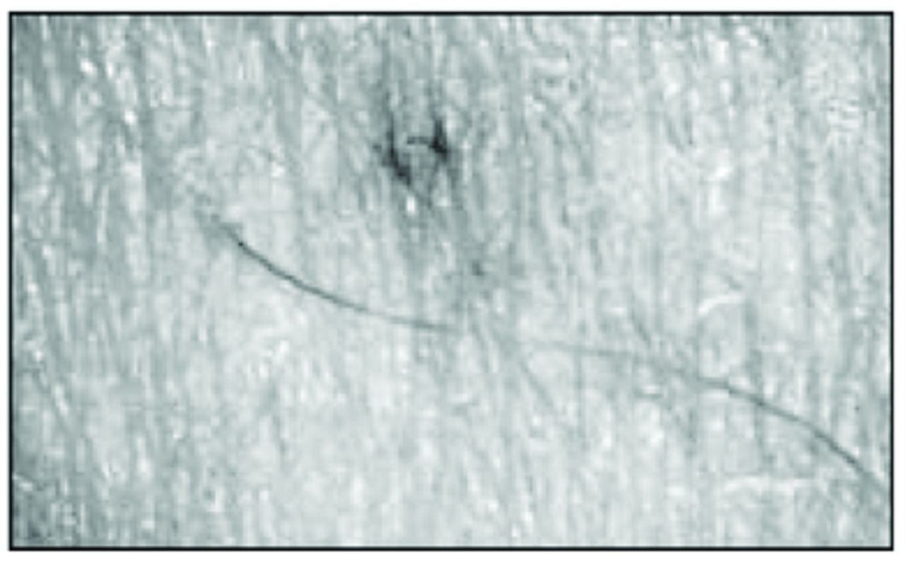
Row II: Lower Segment: Increased perifollicular inflammation (Figure 7)
Row III: Upper Segment: Many inflammatory epithelial cysts (Figure 8)
Row III: Lower Segment: No significant dermal changes
Angle
No consistency of angle
Discussion and Conclusions
This study confirms previous work that production is usually decreased significantly by transection of the follicle.1,2 There is some variability of production depending on the level of bisection1,2,3,4,6 and whether the bisected follicle is part of a two-haired follicular unit.7
Hwang has presented the theory that the recipient area influences greatly the growth characteristics of the hair. Interestingly, the control production rate in this study on the upper thigh was 60%, Hwang’s production on the leg was 60.2%. This compares to a usual 95%+ production on the scalp. Certainly, this study adds credence to Hwang’s theory that Orentreich’s Theory of Donor Dominance is certainly influenced by local growth and reproduction characteristics of the recipient area.10
The rate of growth was significantly diminished. Olsen’s text indicates the average growth rate of scalp hair is 0.37–0.44mm/day.11 This compares to the control growth rate of 2.02mm/month (0.067mm/day), and 1.4 mm/month (0.047mm/day) for bisected hair. Both are significantly diminished compared to those on the scalp. This could have been adversely affected by frictional trauma due to location of this study. The diameter of the controls decreased an average of 4.7um compared to the bisected decrease of 23um. These results certainly confirm but quantify previous observations.1,2,3,4,5,6,8,9
“Because of increased curl, sometimes even to the point of being “kinky,” I have been reluctant to recommend this as a procedure we all should adopt to further refine our hairlines.”
The resultant hairs are lighter in color. It is unknown whether this is because of the decreased caliber of hair shaft giving the illusion of a lighter colored hair or if there is truly a decreased concentration of melanin in the hair follicle.
Swinehart has proposed the use of bisected hairs in the frontal feathering zone.8,9 Because of increased curl, sometimes even to the point of being “kinky,” I have been reluctant to recommend this as a procedure we all should adopt to further refine our hairlines.
There was more perifollicular erythematous epithelial reaction around the transected hair (Figures 7 and 8).


This study would suggest that we should not adopt intentional bisection on a full-scale basis because of the following four reasons:
Decreased survival of the follicle
Increased curl, even to the point of “kinky” hair
Unable to predict consistency of angle because of the shorter segment length
Increased likelihood of erythematous epithelial reaction
The greatest disappointment with this study has been my inability to protect these hairs from local trauma. Because I work out almost every day, there is a lot of frictional trauma in this area of the upper thigh. I would recommend this area not be selected in the future to study hair growth characteristics.
- Copyright © 2002 by The International Society of Hair Restoration Surgery
In this issue


Jump to section
Related Articles
Cited By...
- No citing articles found.