

As hair restoration surgeons, we are all aware of the role of DHT in miniaturization of the hair follicle and of finasteride’s effect in prevention and reversal of this process.
Questions that we should be asking are: How does DHT exert its effect? What processes other than DHT production might we be able to target with new therapies for androgenetic alopecia?
These questions are answered to some extent in the article, “Effects of Finastride on Apoptosis and Regulation of the Human Hair Cycle” (Sawaya ME, Blume-Peyavi V, Mullins D, Nusbaum BP, Whiting D, Nicholson DW, Lotocki G, Keane RW, Eur J Dermatol. 2001;11:304-308).
Apoptosis is a unique mechanism of programmed cell death by which organisms control cell numbers and destroy unwanted cells. Defects in apoptosis can lead to disease pathogenesis, such as in cancer, where there is insufficient apoptosis and cell accumulation occurs or neurodegeneration where excessive apoptosis takes place and cell loss ensues.
A number of studies have provided evidence that apoptosis is a central element in hair follicle regression and is a key player in regulation of the hair cycle, being a prominent feature of the catagen phase.
Apoptosis is mediated by an intracellular proteolytic cascade composed primarily of the caspase family of proteases (Caspase 1-9). These caspase proteases destroy cellular proteins and also activate enzymes that degrade DNA and cellular/nuclear structural proteins eventually resulting in cell death. To avoid inappropiate cell death, there are inhibitors of apoptosis (XIAP, among others) to rescue cells from spurious death signals.
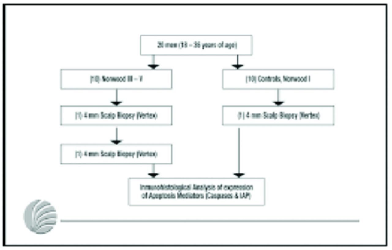
In this study, we used a panel of antibodies and investigated the inmunohistochemical expression of caspases and inhibitors of apoptosis in vertex scalp of Norwood I male patients (“normal” scalp) and in men with AGA before and after 6 months of finasteride 1mg/day (Figure 1).

Study design
Expression of caspases and XIAP was detected predominantly within the isthmic and infundibular hair follicle area, the basal layer of the epidermis, and, to a lesser extent, in eccrine and sebaceous glands. The strongest expression of Caspase-3 (which is the main effector protease) was seen during the catagen phase. AGA-affected tissues showed an increase in caspase immunoreactivity with a concommitant decrease in XIAP staining. After 6 months of finasteride treatment expression of both caspases and XIAP were similar to levels exhibited by normal subjects (Figure 2).

Expressions of Apoptosis mediators in normal subjects and in men with AGA before and after 6 months of finasteride treatment. (Caspase 9 data is from the hair bulb.)
These results suggest that alterations in the levels of caspases and inhibitors of apoptosis play a role in the development of AGA, with increased apoptosis seen in androgenetic tissues as compared to normals. The effects of finasteride on these levels suggest that DHT influences androgen responsive genes that regulate apoptosis in the hair follicle.
These results suggest that alterations in the levels of caspases and inhibitors of apoptosis play a role in the development of AGA, with increased apoptosis seen in androgenetic tissues as compared to normals.
Almost all disease entities have an apoptotic component. There are human clinical data showing anti-tumor responses in melanoma and non-hodgkins lymphoma with oligonucleotide therapy, which affects the RNA of apoptosis inhibitor genes.
Our findings may enhance the development of apoptosis modulating agents to manipulate the hair cycle or treat androgenetic alopecia as well as other hair disorders.
- Copyright © 2002 by The International Society of Hair Restoration Surgery
In this issue


Jump to section
Related Articles
Cited By...
- No citing articles found.