

Methylene blue is a drug widely used in medicine. It is used as a therapeutic drug to treat methemoglobinemia, as an antiseptic to treat infections, and as a temporary skin marker in plastic and dermatologic surgery. It is a very inexpensive material that can be used to facilitate the visualization of premade recipient sites. There are no side effects from absorption since very small amounts are used. Although it is an antiseptic and could be used without previous sterilization, we prefer to do so in order to avoid a rare but not impossible mycobacterium infection.
The steps of the technique are as follows: The recipient area is marked with a regular pen. We divide this area into 1-hair sites, 2-hair sites, and 3-hair sites. After local anesthesia, the lines are tattooed with methylene blue (5%) with a professional tattooing machine (Figure 1). A very slow speed is recommended to avoid a blue line that is too intense. Custom-made blades are used to create sagittal premade sites: 0.75–0.80mm for 1-hair grafts, 1.05–1.20mm for 3-hair grafts, and 0.85–0.95mm for 2-hair grafts.

Lines are tattooed with methylene blue to delineate the recipient area and to separate 1-, 2-, and 3-hair sites.
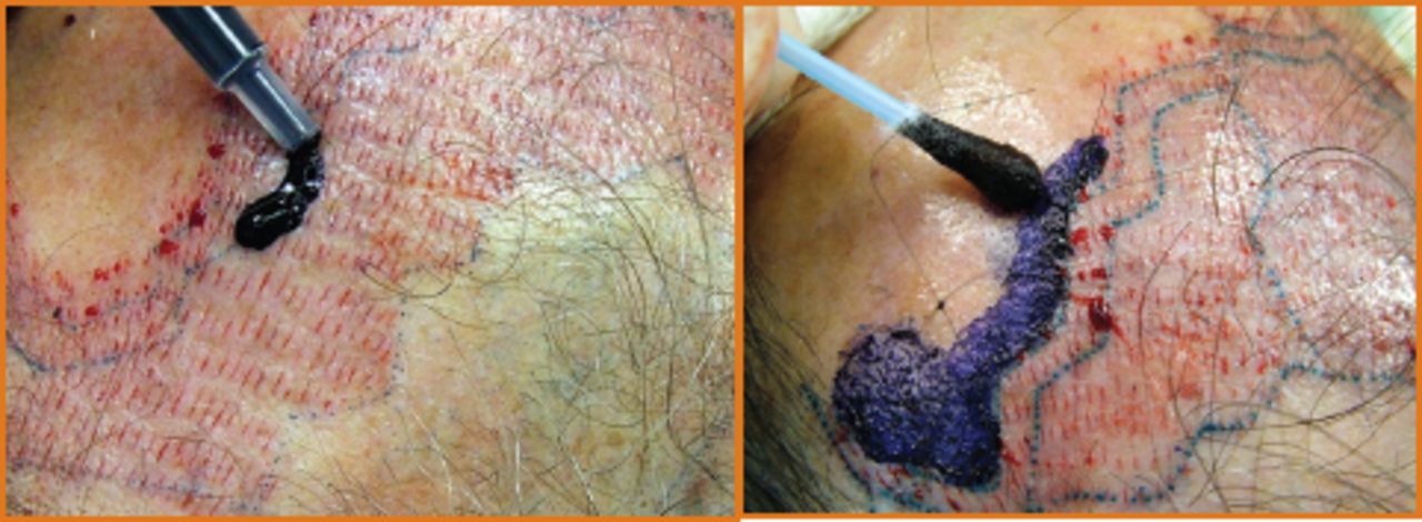
After all the sites are made in approximately 50% to 80% of the recipient area, a few drops of 5% methylene blue are poured over the sites (Figure 2) or applied with a cotton swab. The dye is spread with one finger or a cotton swab, while pulling the skin with the opposite hand to cause traction allowing the methylene blue to penetrate better into the sites (Figure 3). All the sites can be stained or, alternatively, the 1-hair and 3-hair site areas are stained, but not the 2-hair sites, in order to create a contrast among the three areas (Figure 4).

A few drops of 5% methylene blue are poured over the recipient sites with a syringe. It can also be applied with a cotton swab.

Methylene blue is spread with a fi nger or cotton swab while the opposite hand pulls and creates traction on the skin to enhance penetration into the sites.

The 1- and 3-hair sites are stained to help delineate different sized incisions. Alternatively, all sites can be stained. Note the contrast between the stained and unstained areas.
The area is left to dry for about one minute and saline solution is sprayed over the recipient area to clean it. Excessive dye is removed and the tattooed lines and the stained sites remain visible.
After placing the grafts in the stained area, the technicians tell me how many grafts are left. Additional sites are made in the remaining recipient areas and the process is repeated. With better visualization, we speed up the insertion phase of the surgery and avoid difficult-to-find sites that otherwise could remain empty without a graft. We also use this method to separate and identify areas of different site sizes, making it easier to discern between 1-, 2-, or 3-hair sites. Four days post-op, the methylene blue is no longer visible. I use the same technique on all patients and my staff and I are very happy with this new step in our procedure.
Editor’s Note: Recipient site staining using a proprietary gentian violet preparation was first described by Dr. Muhammad Rashid at the Sydney ISHRS Annual Meeting. There is considerable interest among practitioners to determine the optimal method for ease of application as well as which patient characteristics (e.g., dark skin tone) or recipient incision method (e.g., perpendicular or coronal sites) result in the greatest advantage of using this technique. This report describes another material used for this purpose with the additional application of delineating different site sizes.
- Copyright © 2008 by The International Society of Hair Restoration Surgery
References
In this issue


Jump to section
Related Articles
Cited By...
- No citing articles found.