

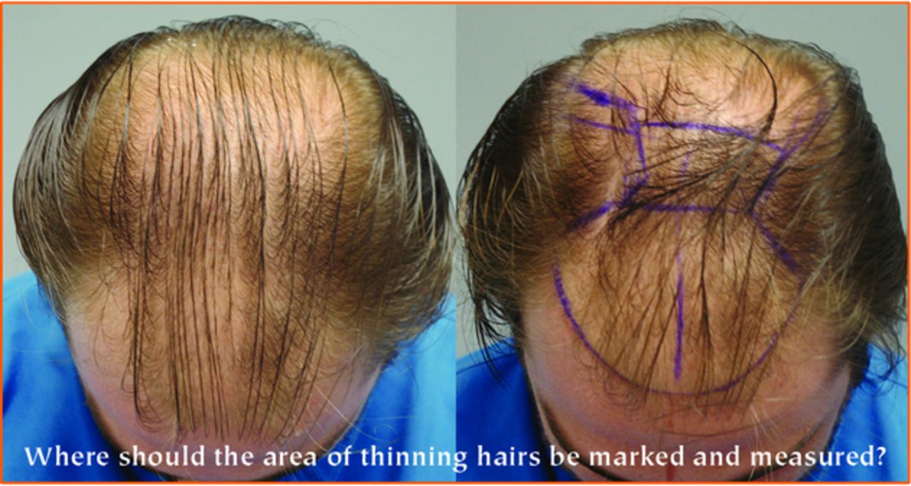
Planning of the recipient area requires an artistic hairline design as well as an accurate outline of the thinning area that needs cosmetic improvement in order for each step of the surgery to be as precise as possible. Based on the total area of coverage, the size of donor area that should be harvested can be decided. From the size of donor area, the number of grafts, which depends on the size of the grafts, can be determined. This sequence is very important for planning the surgery (Figure 1).1
In the past, hair transplant surgeons have used different shaped stencils with predetermined areas to superimpose over the proposed recipient zone. Farjo, et al. suggested the principle of measuring the size of the recipient area by dividing it into simple geometric shapes, such as triangles, rectangles, squares, or circles.2 Cole proposed using the formula for the surface area of an ellipse to measure the total area of the forelock and crown=pi (A)(B), where A is one-half the length and B is one-half the width.3 Farjo further suggested that if only the forelock needs to be measured, then one could simply divide the total of the above calculation by 2.4
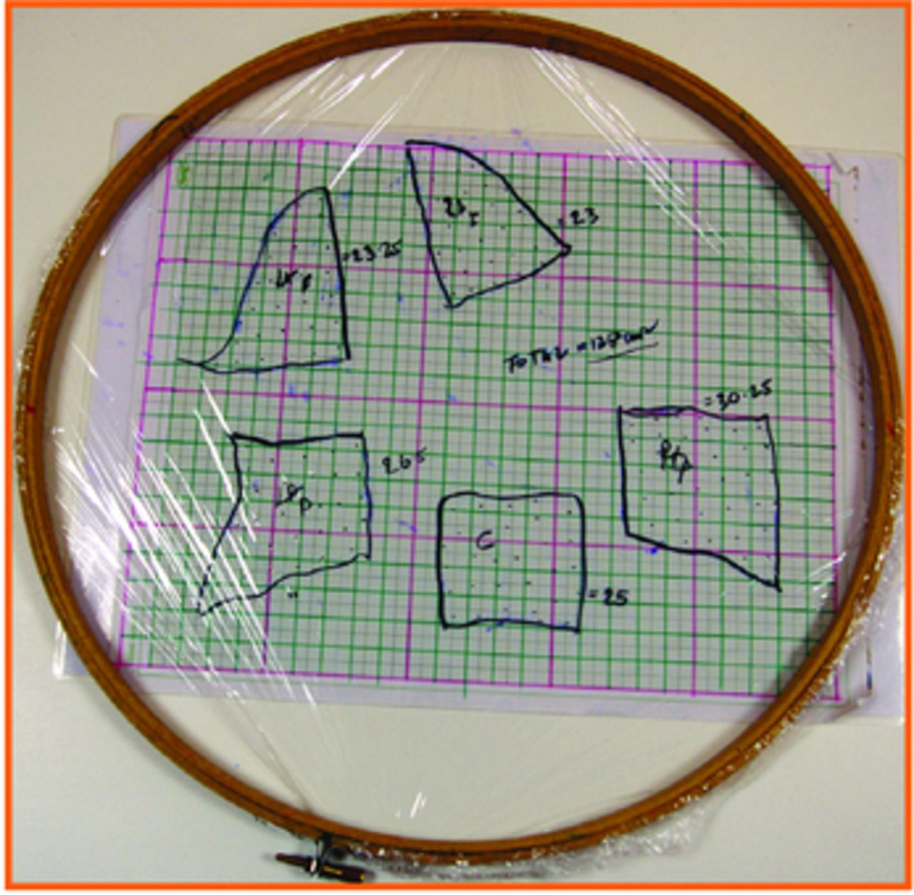
Chang, et al. published the use of a polyurethane wrap (i.e., Saran Wrap) on a circular embroidery ring to trace the recipient area and utilize a 1cm2 grid for the area estimation.5 The method described by Chang is simple and easy to apply.6 One method is to count the intersections in the grid using the principle of morphometrics,5 and the number of intersections will closely approximate the area inside the tracing in centimeters squared.6 However, for a more accurate estimation, counting the actual number of blocks is preferred.6
We have adopted Chang’s method since 2001, however, we have noted some problems in using this method of calculation:
Skin markings are not clearly visible on all skin or hair types, especially with existing hair.
Rocking of the Saran Wrap on the three-dimensional scalp curvature limits the ability to precisely trace the marked line and results in poor reproducibility.
There is inadequate estimation of the traced area via the 1cm2 grid scale, especially at the periphery of the markings.
All these variables led to variations in area calculation among staff members of our clinic.
It is important that the area estimation be valid, be as precise as possible, and be reproducible at all times. For example, a slight difference of 5cm2 (especially if we are planning dense packing with 50 grafts per square centimeter) could make a difference in estimation of 250 grafts.
To develop an efficient, accurate, and reproducible methodology for scalp recipient area measurement, we have refined Chang’s method and compared results with the existing methods of area estimation. Our results were displayed during the free abstract paper presentation at the ISHRS Regional Live Surgery Workshop in June 2010 in Bangkok, Thailand.
Material and Methods
We randomly selected 71 patients who consulted the clinic for consultation or pre-operative assessment.
The estimation for the recipient area was performed using our proposed methodology, which was a refined Chang’s method, Chang’s method, and Farjo’s method. Estimation was also done using either loose Saran Wrap or a transparent shower cap for tracing the markings.
Steps of Our Proposed Methodology (Refined Chang’s Method)
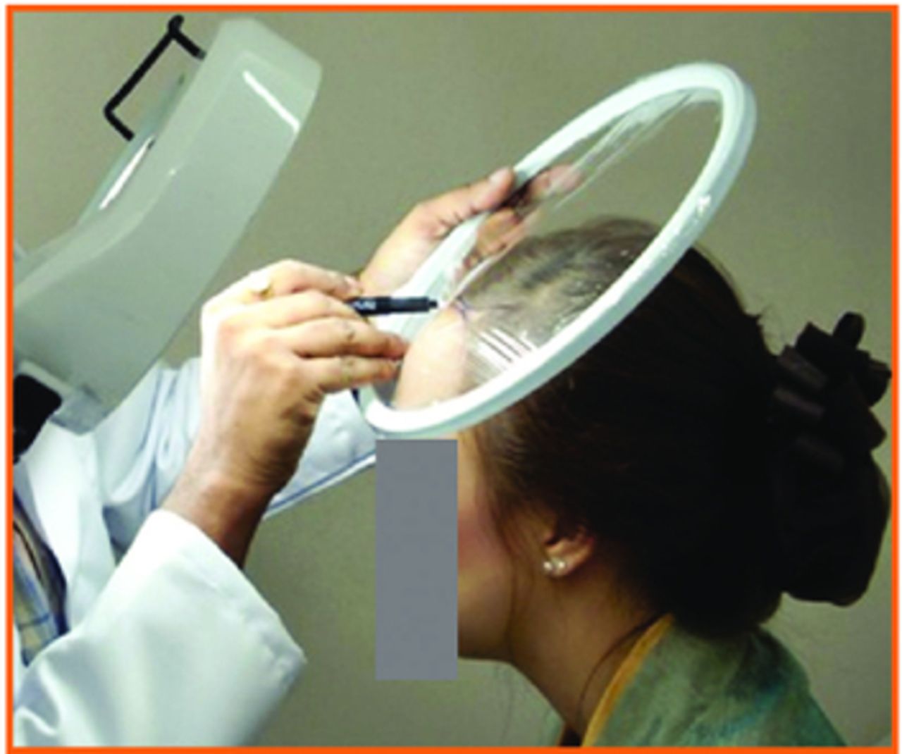
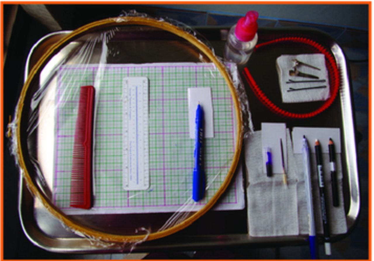
1. Clear visualization of area of baldness. For the assessment of the recipient area, all the patients were examined with bright illumination with or without magnification, and patients’ hair was wet with normal saline or distilled water for better visualization of the thinning area. A hair band and hair clips were used to hold existing hair out of the visual field (Figures 2 and 3).


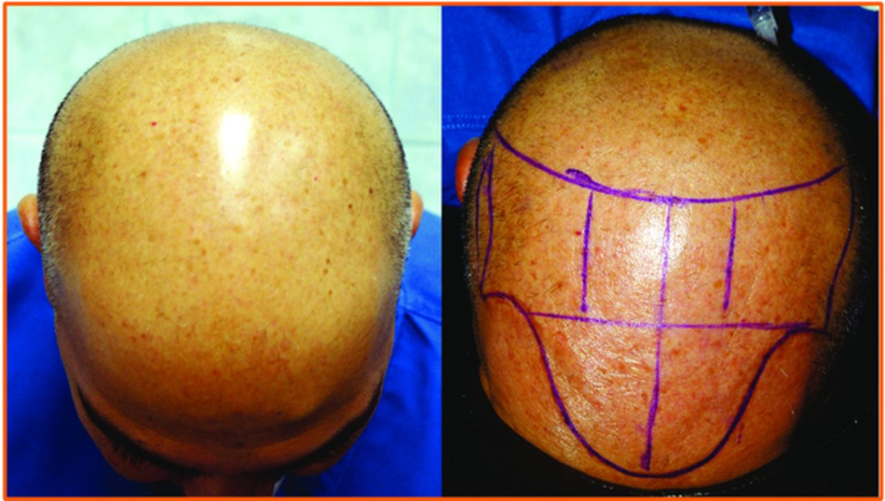
2. Skin marking. The borders of the area of baldness were marked with a finely pointed marker. We used white eyeliner on dark complexioned patients, gentian violent on light skin patients with black hair, and gentian violet or black eyeliner for patients with light skin and blond or grey hair; permanent markers were not used (Figure 4).
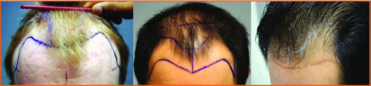
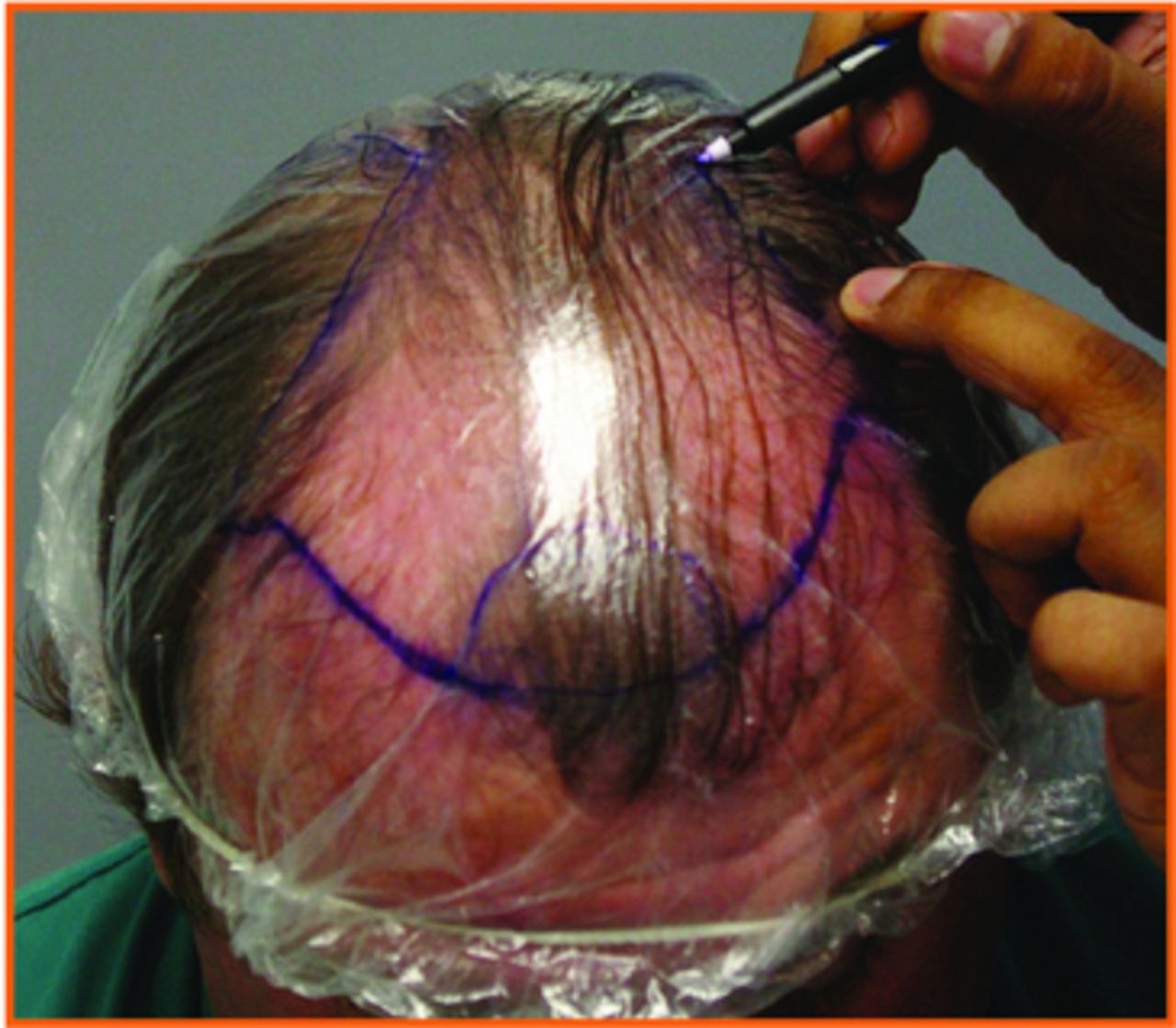
3. Zoning and tracing. For tracing the area of baldness over Saran Wrap, the bald area was divided into small zones wherever the curvature of the scalp changed sharply. Each zone was traced separately on the Saran Wrap (Figures 4 and 5). The individual quadrants are traced separately as described by Chang but without rocking of the saran wrap on the head.

The individual quadrants are traced separately as described by Chang but without rocking of the Saran Wrap on the head.
4. Grid for area calculation. Mathematical graph paper having a grid of 0.25cm2 was used. The total number of small (0.25cm2), medium (1cm2), and big (25cm2) boxes in each zone were counted and the area was estimated (Figure 5). To calculate the total area, the sum from each tracing was calculated. We always try putting the Saran Wrap tracing over the grid scale as tightly as possible with minimal pleating.
For the first 37 patients, we measured the area using the proposed methodology and Chang’s method. For 7 cases, loose Saran Wrap and the shower cap method was used (Figure 7). The results were recorded and compared. The area measurement for another 36 patients was done using all three methods. The method of hair examination and skin marking in all methods was the same, only the tracing method and the calculation grid was specific to the method used. One case was rejected because all three methods could not be used. This case required an irregular area of transplant correction, so we were unable to simulate the irregular area into any shape for area calculation via Farjo’s method. All care was taken to not repeat the recording of cases in whom both consultation and pre-operative assessment was done. All the decimal values were rounded to the closest number and the results were recorded in a Microsoft Excel sheet and analyzed.

Results
Patient demographics are shown in Table 1. Sixty-five patients were estimated using the refined Chang’s method (our proposed methodology), of which 62 cases were compared with the standard Chang’s method, 33 were also compared with Farjo’s method, and 3 cases were compared with the loose Saran Wrap method. In 5 cases, the area measurement was compared between Chang’s method and the shower cap method (Table 2).
On comparing our proposed methodology with the standard Chang’s method, we found an average of an additional 9.23% area measured with the Chang’s method, with the difference ranging from 1.9-24.3%. An average of 17.4% additional area was measured with the shower cap method, ranging from 10-30%. On comparing our refined Chang’s method with Farjo’s method, an excess measurement of 5.12% was found with the latter. Using the loose Saran Wrap, the area measurement was found to be similar to our method (Table 2). We also superimposed the cut tracing back to the scalp marking, which was found to be an exact match using the proposed methodology but not with any other method.
Discussion
Rassman proposed the concept of “multi-variant” analysis for the assessment of the number of grafts required for the cosmetic fullness of hairs on the scalp with 7 variables: color contrast of hair and skin; hair shaft thickness; hair character; size of the bald area; donor hair density; patient expectations; and the available donor supply that will impact the analysis. He also proposed the corrective aesthetic multiplier for four of the seven variables.6,7 The area of baldness is the multiplier that is most variable from doctor to doctor, which leads to inconsistent assessment during planning of the hair transplant surgery session.
In our study, the proposed methodology of area estimation minimizes pitfalls in all aspects of area estimation. For enhancing better visualization, we recommend that hair be wet and higher magnification be used under good light. Sarifakioglu, et al. proposed plastic surgeons require skin markers to have a very fine tip.8 He also asserted that for dark-skinned people, light-colored ink materials (white, green, yellow, red) are more visible.9 Thus, for better color contrast, we advocate use of white eyeliner for dark skin, gentian violet color on patients having white skin with black hair, and gentian violet or black eyeliner for white skin with blond and grey hair. As our proposed method divides the entire recipient area into zones wherever the scalp curvature is sharp, we are able to eliminate the error produced by rocking the Saran Wrap, which makes you lose your tracing. The grid scale of 0.25cm2 area used also enhances accuracy.
We also reevaluated the proposed method by putting back the cut sheet of traced area to the respective zone on the scalp and found it to match while none of the other methods matched. Below are a few of the interesting findings that we observed while comparing the three methods:
With Chang’s method, tracing the marked line on a Norwood Class V or higher patient results in rocking the Saran Wrap from one side to the back and then to another side of the head, thus, it is easy to lose your place in the tracing and add a lot of area in estimation. Near the hairline or on the flat scalp surface, the measurement was found to be almost the same because the Saran Wrap stretches to a flat shape and eliminates the error of rocking.
For measurement of an irregular area, Farjo’s method was found to be very complicated because the zone has to be split into many pieces to conform to geometrical shapes.
For estimation for the frontal area and the hairline with Farjo’s method, using a triangle simulation underestimated the hairline area so we tended to overestimate by simulating the hairline with half of a circle.
Lastly, the hairline height and the hairline design reflect the art and experience of the hair transplant physician, which is always different. This factor leads to differences of area estimation followed by differences in number of grafts required from clinic to clinic.
We experienced that, with the use of Saran Wrap, the pleats may add to the margin of error because stretching it too much to match the scalp three-dimensional surface can result in failure of the wrap to recoil to the normal two-dimensional shape for accurate grid area calculation.
We have also tried to use a transparent shower cap for tracing the area marked on the scalp. Although this is economic, convenient, and fits well to the scalp curvature, the method has the limitation of moderate pleating that adds to the error in area estimation (Figure 7).
- Copyright © 2010 by the International Society of Hair Restoration Surgery
In this issue


Jump to section
Related Articles
Cited By...
- No citing articles found.