

In the previous issue of the Forum, Dr. Cagatay Sezgin from Turkey described an interesting case of donor site necrosis seen after an FUE procedure. We follow with a case submitted by Dr. Kamran Jazayeri from Iran, which describes a well-documented but fortunately rare complication of hair restoration surgery: recipient site necrosis. These types of complications may be distressing both to the surgeon and to the patient, but with appropriate management, positive outcomes may be achieved.
History
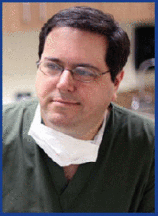
The patient was a healthy 57-year-old man with no significant past medical history except for a 20+ year history of smoking (Figure 1).

57-year-old patient
After consultation, the patient was scheduled for an FUT procedure in order to cover his anterior and midscalp areas. A strip was harvested from the donor area under local anesthesia, with no complications or difficulties. The recipient areas (frontal hairline, front and anterior midscalp) were locally anesthetized with a field block using a 50/50 mixture of 0.5% bupivacaine and 2% lidocaine and 1:100,000 epinephrine. Next, tumescent was prepared by mixing 100ml of normal saline, 40 milligrams of triamcinolone and 1:100,000 epinephrine. A total of 30-40 milliliters of this solution was injected subcutaneously to achieve vasoconstriction and tumescence prior to making the recipient sites.
The recipient sites were made using cut-to-size micro-blades. 0.9mm blades were used for the anterior hairline zone and 1.0- 1.2mm blades for the other areas. The recipient site density varied from 25-35 slits per square centimeter, with the highest density in the frontal core area. All the slits were parallel (sagittal) in orientation. Blades were set to a depth of approximately 4mm, slightly shorter than the length of the patient’s hair follicles. During the operation no abnormal reaction in the recipient area was noticed except for the usual transient blanching due to the effect of the epinephrine.
A total of 1,850 follicular units were transplanted in the frontal hairline, anterior and midscalp areas. The patient returned for bandage removal the day after the surgery with minimal, normal post-op crusting and a normal appearance of the surgical areas.
The patient returned for suture removal 8 days after surgery and at that time an area of thick crusting was noticed in the frontal core (central area) of his anterior scalp (Figure 2). He reported no pain or other symptoms in the affected area. During the next several days he was examined in the office every one or two days and showed no signs of improvement in the crusting, while the surrounding areas appeared normal.

Area of thick crusting noted in the frontal core 8 days’ post-op.
Two weeks after the operation, the adherent eschar was debrided completely using a 3mm curette deep to the subcutaneous layer, and the resulting wound was left open to heal by secondary intention. The patient was advised to clean the wound daily and to apply an antibiotic ointment regularly to the base of the wound. He was also advised to stop smoking.
The debrided wound healed and re-epithelialized in about three weeks, through secondary intention, leaving an atrophic and depressed scar in the central scalp area (Figure 3). Hair growth was achieved in the surrounding areas, but with much less density than what was expected.

Atrophic and depressed scar in the central scalp area.
One year after the first operation, 350 follicular unit grafts were transplanted in the scar and its surrounding areas with a density of approximately 20 follicular units/cm2. No epinephrine was used this time and the patient was strongly advised not to smoke in the days preceding and following the procedure.
The patient did not come back for follow-up so pictures after the revision transplant are not available. However, in a recent telephone call he stated that he is very happy with the final outcome of the procedures.
Discussion
The patient developed an area of full-thickness skin necrosis over the central scalp, behind the hairline. This central area (frontal core) is vascularized with end arteries and is a watershed area. Paradoxically, this is the region where the highest density of grafts is required to achieve a more natural and dense appearance. If the perfusion is compromised, necrosis may occur anywhere in the recipient area, however, the central area is the most vulnerable region.
In patients with risk factors such as atherosclerosis, heavy smoking, and diabetes, the surgeon must use minimal amounts and concentrations of epinephrine in the tumescent solution in order to decrease the risk of vascular perfusion compromise.
Regarding the recipient sites in these patients, the physician should be careful to avoid dense packing and stay on the safe side with a target density below 30 follicular units/cm2 and watch the recipient area carefully for any early signs of perfusion compromise such as blue discoloration (cyanosis).
In my opinion, using needles instead of micro-blades may cause less vascular injury, probably because their sharp, pointed end injures a smaller area in comparison to the rectangular tip of the micro-blades. Minimizing the depth of penetration of the needle may also be helpful.
In the event of necrosis, early detection by closer follow-up and early application of topical nitroglycerin ointment may be helpful. Soon after the limits of the necrotic area are demarcated, debridement of the necrotic eschar and proper wound management to promote secondary intention healing usually leads to complete healing within a few weeks and the resulting alopecic scar may be transplanted later, with some extra precautions.
In order to repair the alopecic scar secondary to the necrosis, it is recommended to wait at least a couple of months to allow for complete healing and revascularization of the area. Also, it is important to counsel the patient about the need to stop smoking. Furthermore, avoiding or limiting the use of epinephrine and preparing the recipient sites with less density, using the smallest needles or blades possible and minimizing the depth of the recipient sites may be helpful.
Columnist’s note from Dr. Marco Barusco: Necrosis of the recipient areas is a rare but well-described complication of hair restoration procedures. It is important to try to identify patients who are at increased risk for poor vascular supply and to do everything possible to try and minimize their intraoperative risk of problems.
Patients at increased risk for recipient area necrosis include:
Older patients
Patients with history of peripheral vascular disease (low perfusion of extremities, history of previous amputations from vascular disease, etc.)
Diabetics under poor control
Patients with history of coronary artery disease and atherosclerosis
Heavy smokers or prolonged history of smoking
Patients with history of sickle cell disease
Patients with history of hypercoagulation (history of deep vein thrombosis, etc.)
Patients previously submitted to scalp flaps, scalp surgeries, or multiple HT procedures
This highlights the importance of a thorough medical history on every patient and a physical exam in case of suspicion (check peripheral capillary refill, peripheral pulses, etc.).
Intra-operatively, the use of high concentration epinephrine for tumescence coupled with high volume of tumescent solution and dense-packing in the central core of the anterior and midscalp may overwhelm the available blood supply to the area, causing ischemia and necrosis. In high-risk patients, central core density should be achieved gradually with multiple procedures instead of with dense packing.
The orientation of the recipient sites seems to also matter, although this is a topic of controversy. Dr. Jerry Wong was asked to give his opinion on the degree of vascular injury and orientation of recipient sites (perpendicular or parallel), and he is of the opinion that perpendicular (coronal) sites cause more injury to the blood supply than parallel (sagittal) ones. I agree with that statement.
Besides the reduction of epinephrine and the volume of tumescence solution used, once an area of persistent blanching or cyanosis is noted, the use of topical nitroglycerin gel may minimize the area of necrosis or avoid it altogether. Care must be taken when using nitroglycerin topically, since it may lower the patient’s systemic blood pressure. A dose of 325mg of aspirin may also be given to the patient, and the application of moist heat to the scalp may promote local vasodilatation and improvement in perfusion.
Once the area of necrosis is delineated and stops progressing, which may take a few days, then debridement and wound care are standard of care. Hyperbaric chambers have been used by some during the healing phase to improve wound closure. Low level laser therapy is also known to improve wound healing and should be considered.
- Copyright © 2014 by The International Society of Hair Restoration Surgery
In this issue


Jump to section
Related Articles
Cited By...
- No citing articles found.